In detecting pathological changes in dentistry, it is important to assess the condition of the teeth based on their appearance.
Ad:
You can read this text in 1 min.
ojoimages
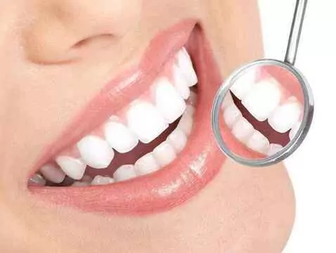
Visit to the dentist


In detecting pathological changes in dentistry, it is important to assess the condition of the teeth based on their appearance.