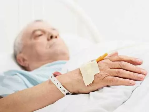
The problem of pressure sores mainly affects immobile patients who spend most of the day in one position. At risk are patients in intensive care units, orthopaedic wards, with multiple injuries, neurological deficits, patients in terminal stages of cancer, cachexia, under hospice care. Due to the severity of the problem of pressure ulcers, the therapeutic difficulties, they require constant care by specialised medical personnel, combined preventive measures and effective, thoughtful therapeutic management.
Pressure sores are ulcer-like lesions - initially confined to the skin, but as they progress, they reach deeper tissues and even muscles or bones. The mechanism of pressure ulcer formation is complex. Local compression of the most adherent tissues is cited as the main cause. This results in occlusion of the blood vessel lumen, local ischaemia, hypoxia and malnutrition of the tissues, reducing their regenerative capacity. In addition, the local reduction in blood flow rate exacerbates the propensity for thrombus formation, intensifying the cascade of local inflammatory and ischaemic response.
Initially, the flow disturbance affects the microcirculation in the superficial layers of the skin. The process observed at this stage can be effectively inhibited by adequate prophylaxis and early local therapeutic measures. If unnoticed early on, local tissue necrosis is initiated in the area of greatest gravitational pressure on the substrate as a result of progressive ischaemia. Difficult-to-heal, deep, penetrating pressure ulcers appear, often succumbing to secondary infection, making effective therapy even more difficult. In addition to pressure, infection and local ischaemia, the pathogenesis of pressure ulcers draws attention to frictional forces that directly damage the delicate skin of the patient.
Local advancement
The classification of pressure ulcers (Torrence scale) divides all pressure ulcer wounds into five categories, according to the degree of progression.
- I - fading redness or erythema of the skin without damage, redness fades under light pressure of the examining finger - microcirculation is not damaged
- II - not fading, increasing redness, superficial maceration of the epidermis, tissues do not yield - erythema does not fade, sometimes swelling of surrounding tissues occurs, usually severe pain is felt
- III - deep damage of the full thickness of the skin up to the border with subcutaneous tissue, surrounded by severe erythema and massive oedema, due to possible nerve damage, it may be less painful, the bottom of the wound filled with yellow masses of disintegrating tissue or red granulation tissue
- IV - the lesion involves subcutaneous tissue, fatty tissue, muscle, even bone, the patient sometimes presents with general symptoms: fever, general malaise.
- V - Advanced penetrating necrosis, deep ulceration occupying the full thickness of the tissues, disintegrating masses of black necrosis.
Predisposition
Not every patient lying down will develop a pressure ulcer at the same time. Of the clinically relevant factors, attention is drawn to the amount and duration of pressure on the skin - but this is individual. In some patients, already a short duration of pressure contributes to the formation of massive, penetrating pressure ulcers.
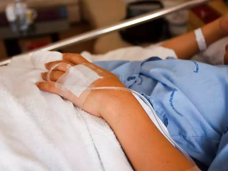
photo: panthermedia
As already mentioned, long-term immobilisation is the main cause of pressure sores. They occur in the areas most exposed to pressure - where bony protrusions lie in close proximity to the skin coverings - so their location varies depending on the patient's body position. In the case of patients lying on their backs, the areas most at risk are the sacrum, iliac crests, heel cusps, elbows, occiput and shoulder blades; in the supine position, the area around the shin, femoral crest, shoulder process or ribs, and the head, depending on the position. The abdominal position is the safest, but this does not mean that the risk is completely eliminated - here they occur mainly on the toes, the external genital area in men, the breast in women, the shoulder process, the sternum, the forehead or the chin. In a sitting position, the sciatic area is at risk; in the event of care errors, they can be located in any region of the body.
Many factors are listed that further potentialise the risk of pressure sores. In neurological patients, with urinary or faecal incontinence, difficulties in maintaining hygiene increase the risk of both the formation and secondary infection of bedsores many times over. Reduced or complete lack of sensation (pain, touch, pressure) impairs the patient's alertness and results in the involuntary formation of pressure ulcers.
Additional chronic diseases predisposing to poorer wound healing are diabetes, atherosclerosis, autoimmune diseases treated with steroids. In addition, poor general condition - cachexia, especially protein-calorie cachexia - or, on the other hand, significant obesity generate conditions for faster ulcer formation. Obese patients often develop pressure sores in the skin folds - these are then difficult to treat and care for locally. Insofar as the patient is found to carry alarm pathogens, there is an increased risk of wound infection and less effective antibiotic therapy.
Tissue damage caused by trauma, also during care procedures, unprofessional positioning of the patient, forcibly moving the body on the ground (rather than transferring) in a way that causes skin and subcutaneous tissue rippling are all factors dependent on the staff caring for the dependent patient.