This article provides a brief historical overview of the surgical treatment of urinary incontinence. It discusses current trends and methods of modern treatment of this condition.
A breakthrough was the use of the endoscopic technique by Vancaillie and Schnessler, a modification of the Burch method of surgery. Laparoscopy, by reducing tissue trauma, contributed to increased patient comfort and a shorter hospitalisation period. The procedure begins with the insertion of an optical trocar and two auxiliary trocars. The wall peritoneum is incised over the filled urinary bladder (approximately 250 ml of fluid). Preparation of the tissues of the załonal space is carried out until access to the pubococcygeal fascia and Cooper's ligaments is gained. We then fixate, using prolene mesh and staplers, the pubocervical fascia and adjacent urethra to the Cooper's ligaments.
Loop operations are typical suspension methods. They aim to elevate the urethrovesical angle through a 'tape' conducted underneath it. There has now been a renaissance of this method.
Stoeckel (1917) was the first gynaecologist to popularise the method of running fascial bands originating from the anterior abdominal wall beyond the pubic conjunctiva and suturing them together below the urethra. He used a modification of the methods developed by Goebell (1910) and Fragenheim (1914).
Other loop techniques used in the treatment of SUI include:
- aldridge surgery,
- Millin,
- Pereira,
- Narik,
- Palmrich.
Research by Petrus and Ulmsten into the mechanisms that ensure urinary continence led to the development of a surgical technique to support the function of the pubovesical ligaments with plastic tape (1996). Today, TVT, IVS and, increasingly, the latest TOT method are the predominant procedures.
In the TVT method, in contrast to the classic method, where the canal is dissected bluntly to guide the tape, special long needles are used here. The tape does not need to be sewn - it remains in the tissue on its own.
The TVT procedure begins with the patient in the gynaecological position with maximum flexion of the lower limbs at the hip joints. The buttocks should be level with the edge of the operating table. For better access, the labia minora may be spread and sutured laterally. A catheter should be inserted into the urethra and the bladder emptied. Above the pubic conjunctiva, two 0.5 cm long incisions are made about 4 cm apart. Approximately 1 cm below the urethra, we incise the vaginal mucosa for a length of approximately 2 cm, which is then dissected laterally.
From the vaginal side, we insert two curved needles consecutively with a prolene tape stretched between them. During the insertion of the needles, the urethra and bladder neck are pushed back to the opposite side using a probe inserted into the Foley catheter. After insertion caudally (just behind the pubic conjunctiva in the lateral direction) of each needle, we perform cystoscopy, checking the continuity of the bladder and urethral walls. We check the fit (adequate tension) of the tape with the hepar (usually No. 8) inserted in the urethra and the tip of the preparation scissors between the urethra and the tape during the cough test. From here, contact with the patient is necessary to induce regional anaesthesia. We then cut off the needles that are brought out over the pubic conjunctiva together with the tape, removing the foil sheath of the tape. The incised skin and vaginal mucosa are sutured.
After the operation, we leave the catheter in the bladder for a period of 2- 5 hours. The operation time is 25 minutes on average. Approximately 80 per cent of patients are completely cured and about 10 per cent of those operated on improve markedly. The most common post-operative complications include urinary retention, the occurrence of urinary tract infections and perforation of the bladder and urethra, and rarely intra-abdominal bleeding.
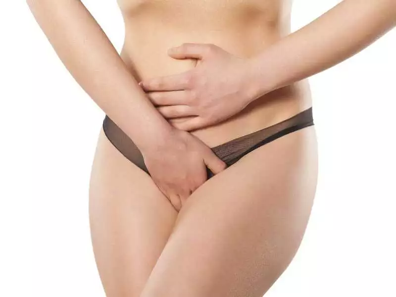
photo: panthermedia
The most recent method using transurethral subcervical tape (TOT) begins with an identical patient positioning and a similar two-centimetre subcervical vaginal incision. We dissect the tissues in lateral directions, up to the inner surface of the ischio-mammary branches, using curved scissors on a blunt "push-and-dilate technique".
#STRONA#
The preparation path of the lateral tunnel should be at a 45-degree angle to the midline, the scissors positioned in a horizontal plane or with the tip pointing slightly upwards. Cut the skin on the inner surfaces of the thighs at the insertion points of the tunnels. To do this, run a horizontal line at the level of the external urethral outlet and then run a second parallel line 2 cm above the first line. The insertion points for the tunnels are on the second line 2 cm lateral to the flexion groove of the thigh (the skin can be flattened by stretching it). We then insert the tunneller without tape from the thigh side into the above skin incision and pierce the obturator opening with a semicircular needle in an outward to inward direction (of the previously dissected periurethral canal) under the control of a finger inserted from the vaginal side. Place one end of the tape over the tunica and pull it in the opposite direction. The procedure is repeated on the other side. The tape is applied without tension and the ends of the tape on the thighs are cut off. Sutures are placed on the skin of the thighs and the vagina, analogous to the TVT.
A feature of this method is the significant reduction in perioperative risks and postoperative complications (bladder and vascular puncture, urinary retention, dysuria). Furthermore, it can be used in an outpatient setting (even under local anaesthesia). The tape used in this method is Uratape (polypropylene, non-elastic tape, silicone-coated in the central-suburethral part) or Obtaje (polypropylene, non-elastic, without silicone coating in the central part).
Other operations used for SUI include:
- extravesical and intravesical bladder neck wrinkling,
- surgery by Louros' method,
- urethral replacement,
- abdominal-vaginal plasty.