


The phenomenon of magnetic resonance was discovered in the 1940s. However, the first MR apparatuses used in clinical examinations did not appear until the early 1980s. MRI is an extremely detailed examination that shows cross-sections of internal organs in all planes. MR examination technology continues to develop and improve - today it is one of the most dynamically evolving disciplines in diagnostic imaging.
Various types of MR systems that use permanent or superconducting magnets are currently used in clinical examinations.
Important! Magnetic resonance imaging (MRI) is excellent for visualising and differentiating individual soft tissues, among other things. MR imaging is one of the most accurate techniques for imaging the brain and can detect abnormalities in the cerebellum, for example.
Characteristics of MR, course of the examination
An MRI is a set of very strong magnets. Despite the high intensity, MRI's magnetic field has no effect on the human body and remains harmless.
Important! MR uses the phenomenon of nuclear magnetic resonance, which makes its invasiveness negligible. Unlike other imaging methods - MRI does not use X-rays, it is based on the natural properties of water molecules in the patient's body.
In clinical applications, a magnetic field in the range of 0, 2 - 3 Tesla is used. The quality of the images obtained increases with the intensity of the applied field and is highest in high-field instruments.
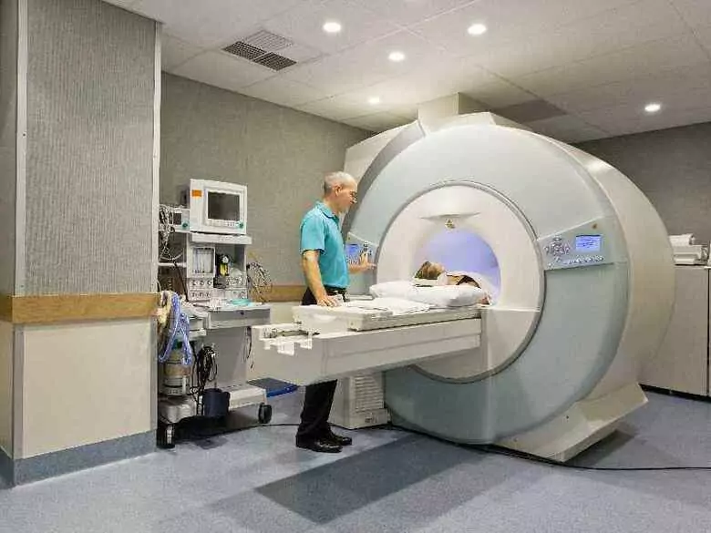
By its design, an MRI resembles a magnet in a shape similar to a long tunnel, which is open at both ends - the patient is placed in the centre. Radio waves create signals, which are then picked up by a receiver in the MR scanner. The signals are processed by a computer processor so that a detailed image of the tissues can be obtained.
The patient is not allowed to drink or eat before the examination.
Important! The MR examination is painless. Only noise may be heard - from time to time the MR scanner makes a tapping sound. The patient will be given special earplugs or headphones to effectively dull any external sounds.
During the examination, the patient is in constant contact with the medical staff, including an intercom.
In some cases, a gadolinium contrast agent is administered intravenously to the patient during the examination, making it easier to obtain a clear view of the part of the body being examined.
Important! Gadolinium is a rare element that exhibits paramagnetic properties on its own. In standard doses, it is not characterised by the possibility of damage to internal organs.
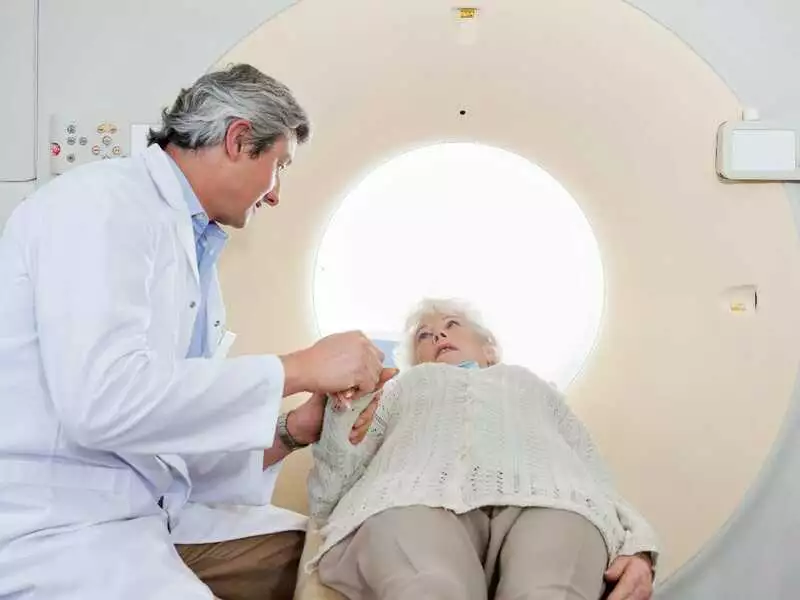
photo: panthermedia
An MR examination usually takes 15 - 45 minutes. It all depends on the area to be examined and the necessary number of images to be obtained. Sometimes the examination can last 60 minutes or longer.
Important! The patient should remain motionless during the examination.
The presence of a strong magnetic field in the MRI causes iron-containing objects to be attracted. It is therefore important to get rid of all metal personal objects (e.g. watch, jewellery) before the examination begins. Women should wash off their make-up (metal micro-particles may be present in cosmetics, which will falsify and interfere with the examination).
Important! In addition, the magnetic field attracts all metal-containing objects in the patient's body (e.g. clips used in the treatment of aneurysms).
In some cases, the MRI examination must be abandoned.