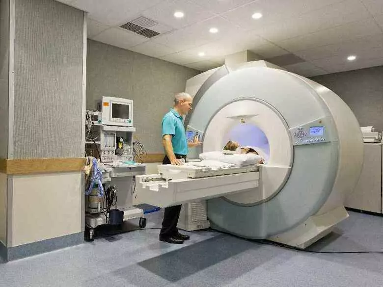


The phenomenon of magnetic resonance was discovered in the 1940s. However, the first MR apparatuses used in clinical examinations did not appear until the early 1980s. MRI is an extremely detailed examination that shows cross-sections of internal organs in all planes. MR examination technology continues to develop and improve - today it is one of the most dynamically evolving disciplines in diagnostic imaging.
Advantages of an MR examination
First and foremost and importantly, the examination does not expose the patient to the negative effects of ionising radiation on the body.
Important! The examination allows the procedure to be performed repeatedly on the same patient.
Other advantages of the MR examination include.
- a painless and non-invasive imaging method that is not associated with exposure to X-rays
- it can be carried out on patients of any age, including pregnant women
- mRI is characterised by a very high spatial resolution, making it possible to visualise fine anatomical details and pathological lesions of very small dimensions
- mRI is the method with the highest contrast capability for soft tissues, which have a different signal (different brightness in the images obtained) in the various MR sequences. This allows excellent differentiation of different tissue types, as well as differential diagnosis of specific types of lesions
- useful in diagnosing a wide range of conditions (e.g. cancer, heart disease)
- facilitates evaluation of both organ structure and function
- detailed images of blood vessels, as well as blood flow, are obtained without having to insert a catheter (no risk of arterial damage)
- provides a rapid, non-invasive method for diagnosing cardiovascular disease
- does not cause any allergic reactions
- allows a non-invasive evaluation of the biliary tract without the need for contrast agent administration
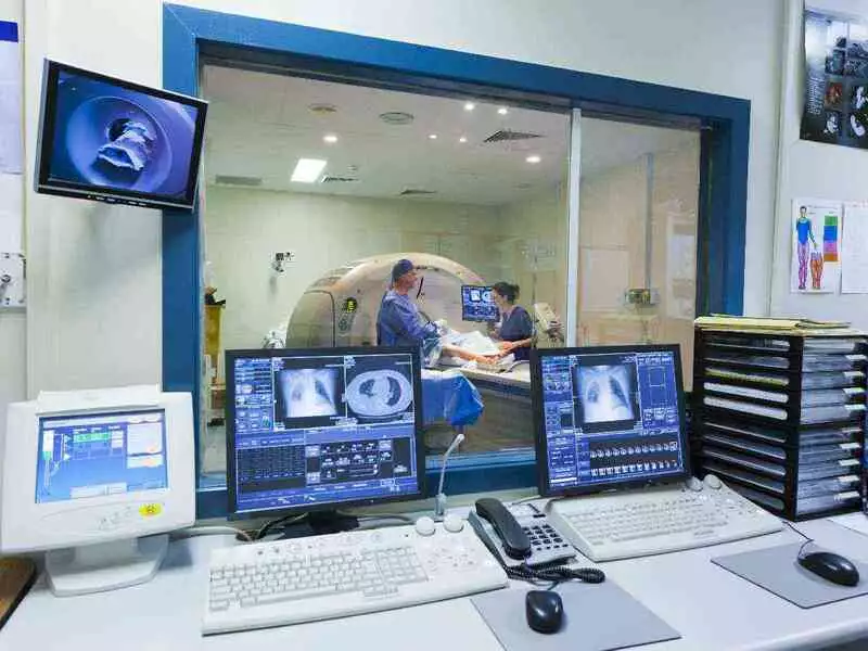
photo: panthermedia
MRI is one of the comprehensive diagnostic methods. The diagnostic scope of MR is extremely wide and includes imaging:
- morphological (structural) imaging of the whole body
- of the urinary tract (MR urography and MR hydrography)
- fluid spaces of the spinal canal (MR pyelography)
- arterial and venous vessels (MR angiography)
- biliary and pancreatic ducts (MR cholangiopancreatography - MRCP)
Another branch of MR imaging is functional imaging - diffusion weighted imaging (DWI), functional MR imaging (fMR), which allows visualisation of brain centres that are responsible for various functions, e.g. the speech centre. Mention should also be made of MR spectroscopy (MRS), a method that allows the chemical composition of the tissues studied to be determined non-invasively.
The images are ready to be described just a few minutes after the examination, making a rapid diagnosis possible. The examination is excellent for trauma patients, young children and pregnant women (no exposure to harmful ionising radiation).
Important! An absolute contraindication preventing MR examination is the presence of implanted electrical devices in the patient's body (e.g. a pacemaker) - the magnetic field present in MR systems can lead to malfunctions of the pacemaker.